Carpets
Cross-Section of a Carpet
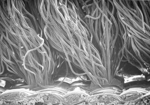
This is a very low power (14x) magnification scanning electron micrograph (SEM) of a cross-section of a piece of carpet that came from the basement of a home. The owner was away on vacation when the air conditioning condensate pump broke, allowing water to soak into part of the two-month-old carpet. When the owners returned, the home and basement smelled musty. Note the lack of any type of particulate matter.
©2020 Jeffrey C. May
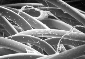
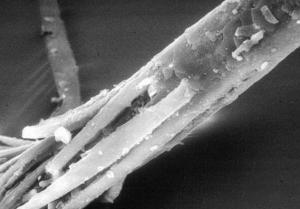
Mold on Carpet Fibers
This is the top of the carpet fibers at higher magnification (150x). At the right and near the top of the SEM, you can see two thin irregular cellulose fibers. The many (larger) smooth tubes are the tops of the nylon carpet fibers. One fiber at the center is covered with the fine, irregular growth of mold hyphae.
©2020 Jeffrey C. May
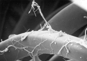
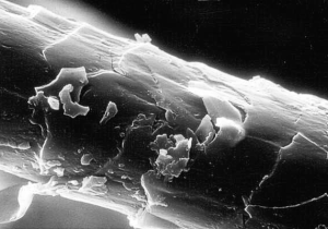
Mold on Carpet Fibers
This SEM at high power (450x) illustrates the mold hyphae wrapped around a nylon carpet fiber (like ivy on a pole) with an aerial projection. The mold is growing on nutrients in the dust settled on the fiber, not on the carpet fiber itself.
©2020 Jeffrey C. May
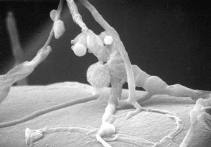
Carpet Fibers
At high power (2000x), this SEM illustrates the base of the aerial projection shown in the previous SEM. Individual round spores can be seen. The rope-like structures are the mold hyphae.
©2020 Jeffrey C. May
Wool Carpet Fiber
This is an SEM (2000x) of the surface of an approximately 27-micron diameter wool carpet fiber from an office. Wool, like many hairs, consists of an interior cortex surrounded by a sheath called the cuticle. The cortex consists of interlocking finger-like tubes with sharp ends, and the cuticle consists of plates, like the scale of a snake. The cuticle of some wool fibers deteriorates, releasing fragments. The smallest fragments in the SEM are about 2 microns; these can become respirable, airborne irritants.
©2020 Jeffrey C. May
Wool Carpet Fiber
An SEM (1000x originally) of a deteriorated wool carpet fiber. The cuticle has fallen away, and the cortex fibers are visible. These fibers can also become airborne irritants when they break up into smaller fragments. The concentration of these particulates above an old wool rug can be hundreds of thousands per cubic meter of air. Symptoms caused by these particulates include eye irritation and coughing. Wool may be itchy because of the physical symptoms associated with the sharp ends of the cortex fibers; exposed cortex fibers at the end of a hair are also responsible for the appearance of “split ends.”
©2020 Jeffrey C. May
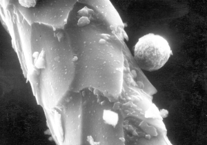
Cat Hair with Paint Sphere
Spheres like this appear in air and dust samples, varying in size from 10 to 70 microns. In a light microscope, they look like circular colonies of bacteria but in reflected light the spheres are the color of the paint, usually white. The bacteria-like particles are titanuam pigment particles. Note the flat portion where another paint sphere was attached at one time. These spheres are produced by spray-painting.
©2020 Jeffrey C. May
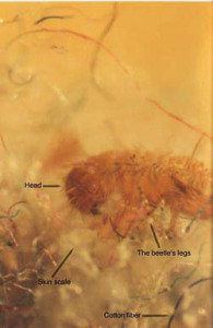
Carpet Beetle
I caught this carpet beetle larva in a vacuumed dust sample from a rug. It is eating a piece of food, possibly a skin scale, surrounded by colored cotton fibers.
©2020 Jeffrey C. May
Ducts
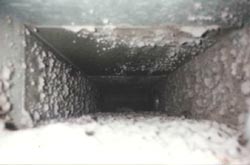
Duct Dust
The inside of a duct before it was cleaned by Rick Hughes (Ductdusters, Winchester, MA). The dust consists mostly of fibers and skin scales. Mites and booklice forage in duct dust while hungry spiders bide their time.
© 1995 Rick Hughes
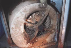
Furnace Blower
A dirty furnace blower before it was cleaned by Rick Hughes (Ductdusters, Winchester, MA). Note the reddish discoloration due to rust. Rusting means that water, probably from a leaking AC condensate pan or furnace humidifier, wet the dust and led to mold growth.
© 1995 Rick Hughes
Mites
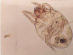
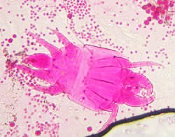
Dust Mite
I trapped this dust mite as it was crawling out of the carpet dust from a very contaminated apartment. Mites are normally quite shy and always hide in dust; this one was escaping from a silverfish that was devouring the mite’s kin. The brown spot at the abdomen is probably a fecal pellet.
©2020 Jeffrey C. May
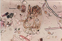
Dust Mite in House Dust
A dust mite, viewed from the bottom side, next to a pine pollen, stained pink; mold and pollen for mites are like candy for children.
©2020 Jeffrey C. May
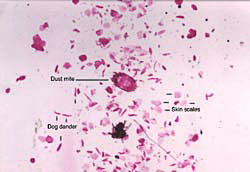
Dust Mite in Air
At the center of this photomicrograph (taken at 100x) is a 140 micron, airborne dust mite (stained pink). At the time I was taking the air sample, a child was playing on a couch, stirring the dust up. The other particles visible are all pink from the acid fuchsin stain and consist almost entirely of human skin scales and dog dander.
©2020 Jeffrey C. May
Mold-eating mite with Aspergillus spores.
Mites like these are commonly found wherever mold is growing indoors, often on stored goods in basements that are not adequately dehumidified.
©2020 Jeffrey C. May
Photographs may be reprinted with permission of May Indoor Air Investigations LLC.